
Key message: When used alone, MTT results can lead to misinterpretation of cellular states in media composition studies. For a more accurate assessment, metabolic assays should be interpreted alongside direct viability measurements, such as Trypan Blue exclusion or other live/dead cell assays.
TThe MTT (3-(4,5-dimethylthiazol-2-yl)-2,5-diphenyl-2H-tetrazolium bromide) assay is widely used to assess cellular metabolic activity and is a well-established tool in toxicology studies. However, because it is cost-effective, simple to perform, and compatible with high-throughput formats, it is also frequently used to infer broader cellular outcomes, including cell viability. Cell viability assays are commonly used to compare how cells respond to different culture conditions. However, when comparing different media supplements, such as fetal bovine serum (FBS) and human platelet lysate (hPL), MTT may not always reflect true cell viability.
This became clear when we compared HEK293T cells cultured under three conditions:
- FBS-supplemented medium
- hPL-supplemented medium
- No supplement control
The aim was to compare cell viability using two commonly used methods: MTT assay and Trypan Blue exclusion (TBE) assay.
What the Two Assays Actually Measure
Although both assays are often described as “viability assays,” they measure different biological properties.
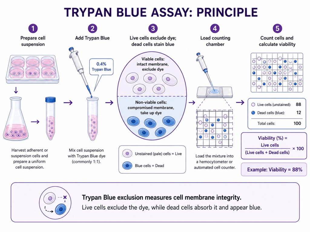
Trypan Blue is a dye exclusion assay. Live cells with intact membranes exclude the dye, while dead cells take it up. Therefore, TBE gives a direct estimate of the percentage of live and dead cells based on membrane integrity.
On the other hand, the MTT assay measures cellular metabolic activity. Viable cells reduce MTT into purple formazan crystals, which can then be quantified by absorbance. A higher MTT signal is often interpreted as higher viability, but this is not always accurate. The signal depends on metabolic activity, mitochondrial/reductive capacity, cell number, proliferation rate, and even the composition of the culture medium.
| Assay | What does it measure | What does it tell you | Main limitation |
| MTT | Metabolic/reductive activity | Metabolic activity is often used as a proxy for viable cells | Cannot distinguish cell number from metabolic activity per cell |
| TBE | Membrane integrity | Percentage of live vs dead cells | May miss early stress/apoptosis if the membrane is still intact |
Experimental Setup
STR-authenticated HEK293T cells were maintained in high-glucose Dulbecco’s Modified Eagle’s Medium (DMEM) supplemented with 2 mM L-glutamine, 4,500 mg/L glucose, 1 mM sodium pyruvate, and 1,500 mg/L sodium bicarbonate. For experimental conditions, cells were cultured in medium containing either 5% human platelet lysate (hPL), 10% fetal bovine serum (FBS), or no supplement as a negative control.
MTT Assay
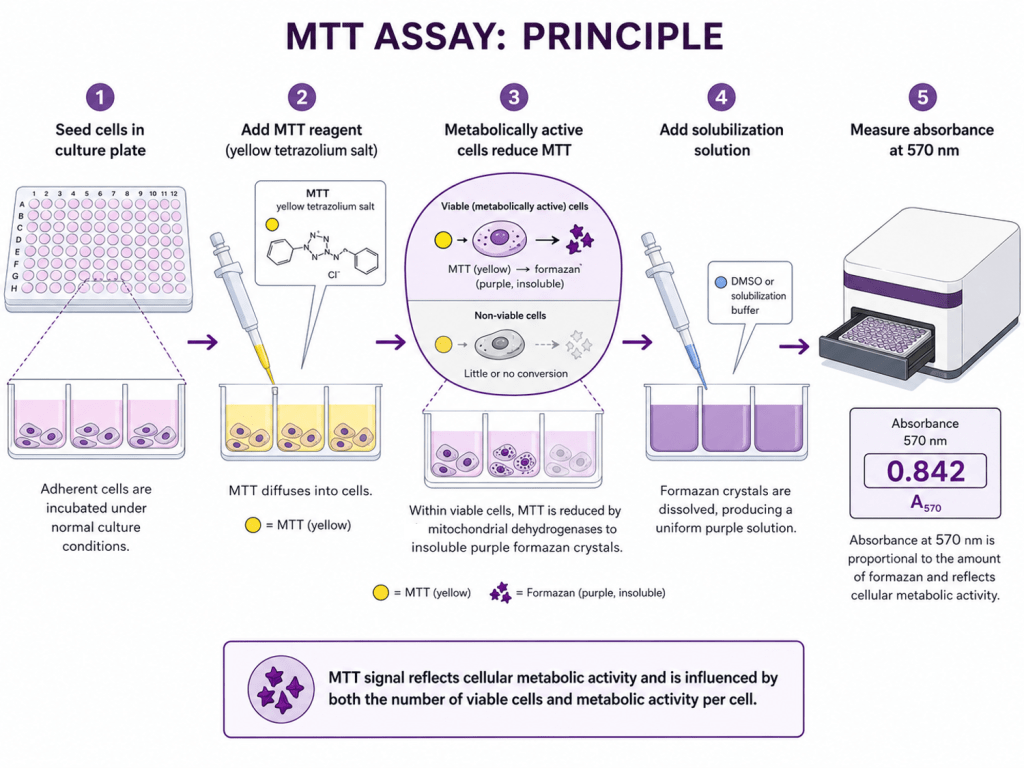
- Seed cells in Culture plate: Cells were seeded in 96-well plates (poly-Lysine treated) at a density of 1×10⁴ cells/well in 100 µl of culture media.
- Add MTT: After 48 h incubation at 37 ℃ (5% CO2), 10 µl of 5 mg/ml MTT stock solution (in PBS) was added to each well (final conc. = 0.5 mg/ml)
- Metabolically active cells reduce MTT: The plate was incubated for 3 h at 37 ℃ (5% CO2). During this time, metabolically active cells convert MTT (yellow) to formazan crystals (purple).
- Add solubilisation solution: Spent media was carefully removed, and 100 µl of filter-sterilized DMSO was added to dissolve the formazan crystals.
- Measure the absorbance: To ensure complete solubilisation, the plate was shaken for 10 minutes using an orbital shaker. The absorbance was measured using a plate reader at 570 nm.
- Data analysis: Absorbance values were considered to be directly proportional to the number of viable cells and normalised to FBS treatment.
TBE Assay
- Prepare cell suspension: Cells were seeded in 24-well plates at a density of 1×10⁴ cells/well in 500 µl of culture media. After 48 h of incubation at 37 °C (5% CO2), cells were washed, trypsinised, and resuspended in 250 µl of PBS to form a uniform cell suspension.
- Add Trypan Blue dye: 0.4% Trypan blue (1:1) was added to the cell suspension, which was then incubated for 2 – 3 minutes at ambient temperature.
- Load counting chamber: 10 µl of the mixture was loaded into a hemocytometer.
- Count cells and calculate viability: The total number of cells and number of viable cells were counted and expressed as a percentage of the live to total cells.
What We Observed
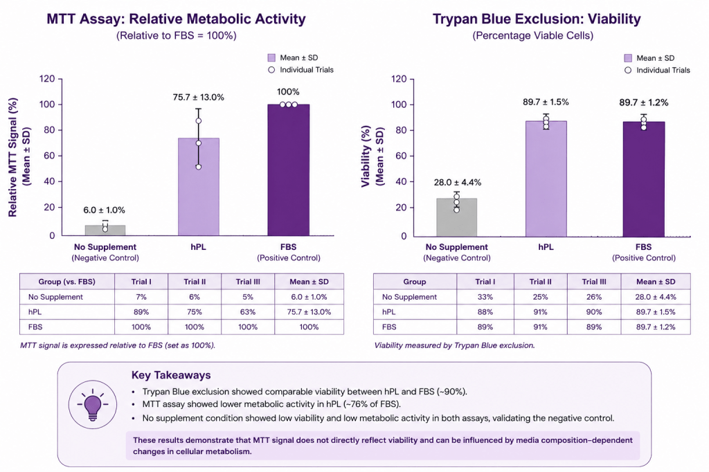
Across three independent trials, TBE showed that hPL and FBS supplementation maintained very similar cell viability. The no-supplement control showed much lower viability, confirming that supplementation was necessary for maintaining cell proliferation and health. On the other hand, the MTT data told a slightly different story. When MTT signal was blank-corrected and expressed relative to FBS, hPL showed a lower metabolic signal across the three trials. These results suggest that using MTT as a proxy for viability can lead to misinterpretation of the effects of media composition in such studies.
Why This Matters
If MTT alone had been used, one might conclude that hPL was less supportive of cell viability than FBS. However, the TBE data do not support that conclusion. Instead, the cells in hPL remained viable at levels comparable to FBS.
In media composition studies, this distinction matters. Different supplements may affect cellular metabolism without necessarily affecting cell survival. hPL and FBS differ in composition, including growth factors, cytokines, proteins, lipids, and other bioactive components. These differences may alter mitochondrial activity, proliferation rate, or cellular reductive capacity, all of which can influence the MTT signal. Therefore, a lower MTT signal does not automatically mean that fewer cells are alive.
Takeaway
Although this study is limited by a small dataset, the findings highlight the need for caution when interpreting MTT results as direct measures of cell viability. For studies comparing culture supplement composition, MTT should not be used alone to conclude cell viability. It is better used alongside a direct viability assay such as TBE, automated live/dead counting, or flow cytometry-based viability staining. A more accurate interpretation comes from combining assays to help distinguish true loss of viability from changes in metabolism.
- Trypan Blue tells us whether cells are alive or dead.
- MTT tells us how metabolically active the cells are.
Further reading
- Ghasemi M, Turnbull T, Sebastian S, Kempson I. The MTT Assay: Utility, Limitations, Pitfalls, and Interpretation in Bulk and Single-Cell Analysis. Int J Mol Sci. 2021 Nov 26;22(23):12827. doi: 10.3390/ijms222312827. PMID: 34884632; PMCID: PMC8657538.
- Stepanenko AA, Dmitrenko VV. Pitfalls of the MTT assay: Direct and off-target effects of inhibitors can result in over/underestimation of cell viability. Gene. 2015 Dec 15;574(2):193-203. doi: 10.1016/j.gene.2015.08.009. Epub 2015 Aug 7. PMID: 26260013.
- Wang P, Henning SM, Heber D. Limitations of MTT and MTS-based assays for measurement of antiproliferative activity of green tea polyphenols. PLoS One. 2010 Apr 16;5(4):e10202. doi: 10.1371/journal.pone.0010202. PMID: 20419137; PMCID: PMC2855713.
 ULQA Scientific
Leave a comment